Muscular dystrophy — dogs take the lead

Duchenne muscular dystrophy (DMD), the most common fatal genetic disease in children, may soon have a cure thanks to groundbreaking US research.
Scientists used CRISPR gene editing to halt DMD in dogs, which, as a large mammal, suggests that a lifesaving treatment for children should follow soon.
The research was conducted by the University of Texas (UT) Southwestern Medical Center, and documents show significant improvement in the muscle fibres of dogs with DMD.
Researchers used a single-cut gene-editing technique to restore dystrophin in muscle and heart tissue by up to 92% of normal levels. Scientists have estimated a 15% threshold is needed to significantly help patients.
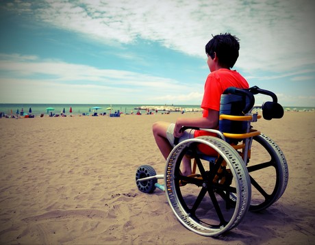
DMD is caused by a mutation that inhibits the production of dystrophin, a protein critical for muscle function.The disease, which affects one in 5000 boys, leads to muscle and heart failure and premature death by the early 30s. Patients are forced into wheelchairs as their muscles degenerate and eventually onto respirators as their diaphragms weaken. No effective treatment exists, though scientists have known for decades that a defect in the dystrophingene causes the condition.
“Children with DMD often die either because their heart loses the strength to pump or their diaphragm becomes too weak to breathe,” said Dr Eric Olson, Director of UT Southwestern’s Hamon Center for Regenerative Science and Medicine. “This encouraging level of dystrophin expression would hopefully prevent that from happening.”
Proof of concept
The study, published in Science journal, establishes the proof of concept for single-cut gene editing in dystrophic muscle and represents a major step towards a clinical trial. Already Dr Olson’s team has corrected DMD mutations in mice and human cells by making single cuts at strategic points of the mutated DNA.
The latest research applied the same technique in four dogs that shared the type of mutation most commonly seen in DMD patients. Scientists used a harmless virus called adeno-associated virus (AAV) to deliver CRISPR gene-editing components to exon 51, one of the 79 exons that comprise the dystrophin gene.
CRISPR edited the exon, and within several weeks the missing protein was restored in muscle tissue throughout the body, including 92% correction in the heart and 58% in the diaphragm, the main muscle needed for breathing.
“Our strategy is different from other therapeutic approaches for DMD because it edits the mutation that causes the disease and restores normal expression of the repaired dystrophin,” said Dr Leonela Amoasii, lead author of the study. “But we have more to do before we can use this clinically.”

The lab will next conduct longer-term studies to measure whether the dystrophin levels remain stable and to ensure the gene edits do not have adverse side effects.
Dr Olson hopes the next step beyond dogs is a clinical trial, which would be among several that UT Southwestern’s gene therapy centre aims to launch in the coming years to address numerous deadly childhood diseases.
Webinar: Turning data protection into a business advantage
Backup is no longer a safety net, but a strategic tool for risk reduction — and the...
Report explores Australian perceptions of Medtech access
A nationally representative survey conducted from August–September 2025 has explored...
Updated guidance for telehealth and virtual care now available
Reflecting concerns around unethical practice and emerging business models focused more on profit...


![[New Zealand] Transform from Security Awareness to a Security Culture: A Vital Shift for SMB Healthcare — Webinar](https://d1v1e13ebw3o15.cloudfront.net/data/89856/wfmedia_thumb/..jpg)
![[Australia] Transform from Security Awareness to a Security Culture: A Vital Shift for SMB Healthcare — Webinar](https://d1v1e13ebw3o15.cloudfront.net/data/89855/wfmedia_thumb/..jpg)


