'Mini-guts' offer clues to paediatric GI illness

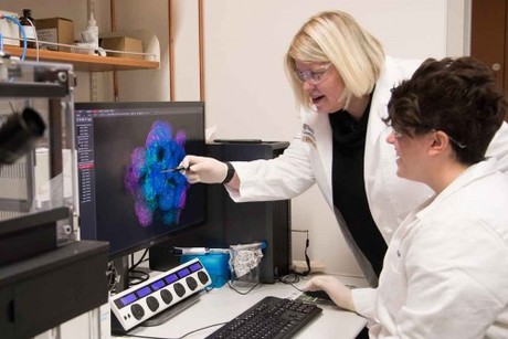
Immature stem cells have been used researchers at Washington University School of Medicine in St. Louis and the University of Pittsburgh to create a miniature model of the gut in the laboratory. The study, published in the Proceedings of the National Academy of Sciences, demonstrates how infection-causing enteroviruses enter the intestine.
Enteroviruses cause millions of infections each year worldwide and can spur flu-like symptoms or more serious conditions such as inflammation in the brain or heart, acute paralysis or even death. Enterovirus infections within neonatal intensive care units (NICU) can be particularly devastating with infant fatality rates approaching 20%.
“This study not only provides important insights into enterovirus infections but also provides an important model that could be used to test the efficacy of antienterovirus therapeutics in the premature intestine,” said Misty Good, MD, an assistant professor of paediatrics at Washington University School of Medicine in St. Louis and co-senior author of the study.
Researchers isolated stem cells from a premature infant’s small intestine and created ‘mini-guts’ in the laboratory as a model to see how the virus behaves in the intestine. This model replicates various cell types within the tissue found in the intestine.
The mini-gut model shows that the small intestines are most susceptible to infection by certain enteroviruses such as echovirus 11 (E11) and coxsackievirus B (CVB), both of which are commonly associated with illness in the neonatal population. E11, the enterovirus most commonly associated with NICU infections, induced significant damage to the mini-guts.
Researchers also found that E11 targets certain cells within the gastrointestinal tract and may allow passage of the virus into the bloodstream, which can result in serious illness.
“Despite their major global impact, especially on the health of children, little is known about the route that these viruses take to cross the intestine, their primary point of entry,” said co-senior author Carolyn Coyne, PhD, an associate professor of microbiology and molecular genetics at the University of Pittsburgh School of Medicine. “Our approach has for the first time shed some light on this process. This model also could be used for developing antienterovirus therapeutics targeting the gastrointestinal tract, given that no therapeutic approaches exist to combat infections of these viruses.”
Said Gary A Silverman, MD, PhD, the Harriet B. Spoehrer Professor and the head of the department of paediatrics at Washington University: “This research has the potential for a positive global impact on human health, especially since enteroviruses cause disease in all human populations and age groups.”
Medicine shortages are testing Australia's health system and clinicians need national support
Medicine shortages are no longer isolated supply disruptions. They are escalating and directly...
Mindfulness and endoscopies — a more effective combination?
A UK study involving 231 patients has advocated 'mindful endoscopy' — allowing...
How do different types of pain influence empathy?
Different types of pain influence how unpleasantly we perceive it but also how we empathise with...


![[New Zealand] Transform from Security Awareness to a Security Culture: A Vital Shift for SMB Healthcare — Webinar](https://d1v1e13ebw3o15.cloudfront.net/data/89856/wfmedia_thumb/..jpg)
![[Australia] Transform from Security Awareness to a Security Culture: A Vital Shift for SMB Healthcare — Webinar](https://d1v1e13ebw3o15.cloudfront.net/data/89855/wfmedia_thumb/..jpg)


